
Fig. 2
- ID
- ZDB-FIG-111031-3
- Publication
- Kroehne et al., 2011 - Regeneration of the adult zebrafish brain from neurogenic radial glia-type progenitors
- Other Figures
- All Figure Page
- Back to All Figure Page
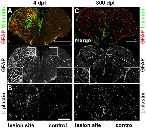
Reactive gliosis, inflammatory response and scarring. (A) The glial markers GFAP (red) and vimentin (green) are strongly upregulated in the lesioned hemisphere early after injury (4 dpl). GFAP+ radial fibres in the lesioned hemisphere are thicker compared with the control hemisphere. (B) The pan-leukocyte marker L-plastin is strongly upregulated in the lesioned hemisphere at 4 dpl. (C) No difference in number and appearance of GFAP+ (red) radial processes is detected comparing lesioned and control hemispheres 300 dpl. Staining for L-plastin (green) shows no difference in the number and distribution of leukocytes between the lesioned and the control hemisphere. Scale bars: 200 μm. |