
Fig. 1
- ID
- ZDB-FIG-111031-2
- Publication
- Kroehne et al., 2011 - Regeneration of the adult zebrafish brain from neurogenic radial glia-type progenitors
- Other Figures
- All Figure Page
- Back to All Figure Page
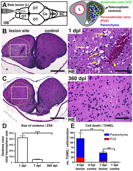
The stab lesion assay and restoration of tissue integrity of the adult telencephalon. (A) Insertion of a canula into the telencephalon of an adult zebrafish causes a lesion canal (L) in parallel to the rostrocaudal body axis along one hemisphere (dorsal view; OB, olfactory bulb; DT, dorsal telencephalon; OT, optic tectum: Ce, cerebellum; SC, spinal cord). In cross-sections (right), three layers can be functionally distinguished in DT: contacting the ventricle is the neural progenitor containing ventricular zone (VZ, green). During constitutive neurogenesis, all newborn neurons integrate into the periventricular zone (PVZ, red, one or two cell diameters adjacent to the VZ). The uninjured central parenchyma (blue) does not receive new neurons. (B) Tissue within the stab canal is strongly vacuolated (arrows), and often contains a prominent blood clot (arrowhead) as seen in Haematoxylin/Eosin (HE) stained sections 1 day post lesion (dpl). (C) Complete morphological restoration of the tissue architecture is seen by 360 dpl. Scale bars: 200 μm in B,C overviews; 50 μm in B,C insets. Circles indicate the lesion canal. (D) Quantification of the size of the vacuolated zona status spongiosus (ZSS) at 1, 7 and 360 dpl (n=3, n=4 and n=3, respectively). (E) Quantification of TUNEL+ cells at 4 hours post lesion (hpl) (n=3) and at 1 dpl (n=4). Data are mean+s.e.m. ***Pd0.001; **Pd0.01. |