
Fig. S10
- ID
- ZDB-FIG-111031-16
- Publication
- Kroehne et al., 2011 - Regeneration of the adult zebrafish brain from neurogenic radial glia-type progenitors
- Other Figures
- All Figure Page
- Back to All Figure Page
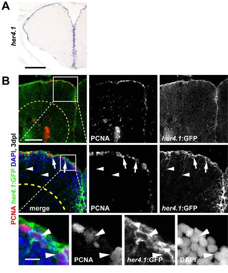
her4.1 is expressed by radial glia showing reactive proliferation after stab lesion. These cells can be lineage traced using tg(her4.1:mcherryT2ACreERT2) transgenic fish. (A) her4.1 is expressed along the whole extent of the ventricular proliferation zone of the telencephalon, as shown by in situ hybridisation. (B) her4.1-expressing radial glia in the VZ react to lesion and re-enter the cell cycle. After stab lesion, tg(her4.1:EGFP) transgenic fish many nuclei of EGFP+ (GFP) cells (green) co-localize with PCNA (red, arrows) 3 dpl, as shown in single confocal sections (high magnification inset). Note that EGFP expression in this line highly mimics radial glia morphology (radial processes, arrowheads). Scale bars: 200 μm in A; 100 μm in B, 10 μm in inset (high magnification). Dashed outlines represent the lesion. |