
FIGURE
Fig. S5
- ID
- ZDB-FIG-111031-12
- Publication
- Kroehne et al., 2011 - Regeneration of the adult zebrafish brain from neurogenic radial glia-type progenitors
- Other Figures
- All Figure Page
- Back to All Figure Page
Fig. S5
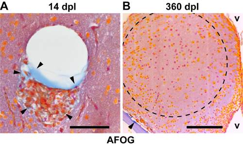
Collagen transiently accumulates at the lesion site. (A) 14 dpl cells at the lesion site are associated with ectopic extracellular matrix (arrowheads, collagen, blue), as seen in Acid-Fuchsin-OrangeG (AFOG) stained 1 µm paraffin sections. (B) At 360 dpl, no ectopic accumulation of collagen is seen in the lesioned hemisphere, indicating absence of fibrotic scar tissue. The collagen-containing bone of the skull (floor plate) is clearly stained blue (arrowhead). v, ventricle. Scale bars: 50 μm in A; 100 μm in B. Dashed outline represents the lesion canal. |
Expression Data
Expression Detail
Antibody Labeling
Phenotype Data
Phenotype Detail
Acknowledgments
This image is the copyrighted work of the attributed author or publisher, and
ZFIN has permission only to display this image to its users.
Additional permissions should be obtained from the applicable author or publisher of the image.
Full text @ Development