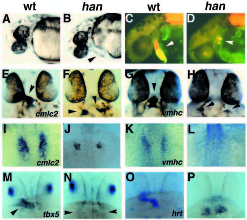
Myocardial defects in hans6 mutants. (A,C,E,G,I,K,M,O) Wild-type embryos; (B,D,F,H,J,L,N,P) hans6 mutant siblings. (A-D) Lateral views at 36 hpf, anterior to the left. (A,B) Bright-field images; mutants (B) display mild pericardial edema (arrowhead). (C,D) Immunofluorescent images of embryos stained with MF20 (TRITC) and S46 (FITC). In these double exposures, red fluorescence indicates MF20 staining of ventricular and somitic tissue, while yellow fluorescence indicates the overlap of S46 and MF20 staining in atrial tissue (Stainier and Fishman, 1992). (C) Wild-type embryos have a midline heart tube (arrowhead) with two distinct chambers, an anterior ventricle (red) and a posterior atrium (yellow). (D) hans6 mutants have two small clusters of myocardial tissue (arrowhead) that appear to be primarily atrial (yellow). (E-H, M-P) Dorsal views through the head at 33 hpf, anterior to the top. (M-P) are golden homozygotes. (I-L) Dorsal views of the myocardial precursors at the 15-somite stage (16.5 hpf). (E,F,I,J) In situ hybridization showing expression of the myocardial marker cmlc2 (Yelon et al., 1999). (E) Wildtype embryos express cmlc2 throughout the heart tube (arrowhead); (F) hans6 mutants have two small patches of cmlc2-expressing myocardial tissue (arrowheads). Younger wild-type embryos (I) have more myocardial precursors than hans6 mutant siblings (J). (G,H,K,L) Expression of the ventricular marker vmhc (Yelon et al., 1999). Wildtype embryos (G) express vmhc only within the future ventricle (arrowhead). hans6 mutants vary in amount of vmhc-expressing tissue: some hans6 mutants have no vmhc expression at this stage (data not shown), while others (H) have small populations of cells with weak vmhc expression (arrowheads). (K) Younger wild-type embryos express vmhc in a medial subset of myocardial precursors (Yelon et al., 1999); (L) vmhc expression is difficult to detect in hans6 mutants at this stage. (M,N) Expression of tbx5 in dorsal retina and heart tube (arrowhead) is apparent in wild-type embryos (M), but only dorsal retina expression is detectable in hans6 mutants (N, arrowheads indicate location of myocardium). (O,P) Expression of hrt in myocardium is apparent in wild-type embryos (O) and in hans6 mutants (P).
|
