
Fig. 3
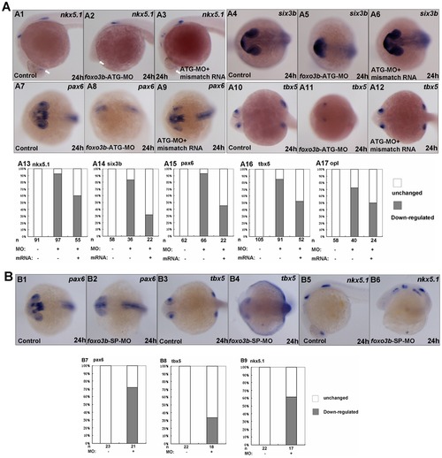
Loss of foxo3b function results in anterior defects. (A) The foxo3b-ATG-MO injected embryos showed remarkable loss of expression of anterior neural markers by 24 hpf, which could partially be rescued by co-injection of foxo3b mismatch mRNA. (A1–A3, A13) The expression of nkx5.1 at the telencephalon (indicated by white arrows) was dramatically reduced in foxo3b-ATG-MO injected embryos. Co-injection of foxo3b mismatch mRNA could partially restore its expression at the telencephalon. (A4-A6, A14) Six3b was specifically expressed at the telencephalon and eyes. Loss of foxo3b function resulted in abnormal expression pattern of six3b, which was restored by co-injection of foxo3b mismatch mRNA. (A7–A9, A15) Pax6 expression at the forebrain and eyes decreased greatly in foxo3b morphants. Co-injection of foxo3b mismatch mRNA partially restored its expression. (A10–A12, A16) Expression of tbx5 at the retina was dramatically reduced in foxo3b-knockdown embryos. Its expression was rescued by co-injection of foxo3b mismatch mRNA. (A17) Opl expression at the telencephalon was reduced in foxo3b morphants compared to control embryos, which was efficiently rescued by co-injection of foxo3b mismatch mRNA. Embryos were injected with 8 ng foxo3b-ATG-MO or 125 pg foxo3b mismatch mRNA,wild-type embryos were used as control. A1-A3, lateral views with anterior to the left; A4-A12, dorsal views with anterior to the left; A1-A17, 24 hpf. (B) The foxo3b-SP-MO injected embryos exhibited anterior defects similar to that of foxo3b-ATG-MO injected embryos. (B1–B2, B7) The expression of pax6 at the telencephalon and eyes was reduced in foxo3b-SP-MO injected embryos. (B3–B4, B8) Tbx5 expression at the eyes decreased in foxo3b-knockdown embryos. (B5–B6, B9) Loss of zygotic foxo3b function resulted in reduction of nkx5.1 expression at the telencephalon. Embryos were injected with 16 ng STD-MO (control) or 16 ng foxo3b-splice-MO. B1–B4, dorsal views with anterior to the left; B5-B6, lateral views with anterior to the left; B1-B9, 24 hpf. |
| Genes: | |
|---|---|
| Fish: | |
| Knockdown Reagents: | |
| Anatomical Terms: | |
| Stage: | Prim-5 |
| Fish: | |
|---|---|
| Knockdown Reagents: | |
| Observed In: | |
| Stage: | Prim-5 |