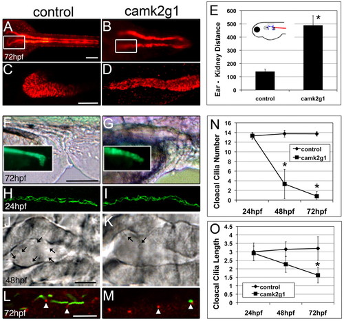
Suppression of γ1 CaMK-II (camk2g1) by injection of the camk2g1 MO (1ng) induces anterior and posterior kidney defects. (A,B) Dorsal view of entire pronephric ducts immunostained for α1 Na+/K+-ATPase; (C,D) regions outlined by white boxes are shown in z-stack projections. (E) Anterior migration is blocked in camk2g1 morphants as inferred from the increase in the distance between the anterior kidney and posterior ear. (F,G) Morphogenic alterations in the ductal region of the pronephros are evident using both DIC optics and GFP-α1 Na+/K+-ATPase fluorescence (insets). (H,I) Pronephric ductal cilia appear normal at all time points as shown by acetylated tubulin immunostaining at 24 hpf. (J,K) DIC images of the cloaca at 48 hpf. Arrows indicate cilia. (L,M) Acetylated tubulin (green) and γ-tubulin (red) show a loss of cloacal cilia but a retention of the basal body (arrowheads) in morphants. (N,O) Cloacal cilia number and length (of remaining cilia) were measured at 24, 48 and 72 hpf. *P<0.005. Scale bars: 100 μm in A,C,F; 5 μm in J,L.
|
