
Fig. 3
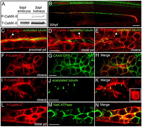
Activated CaMK-II is enriched in the zebrafish pronephric kidney. (A) Immunoblot of P-CaMK-II and total CaMK-II in whole embryo (6 dpf) and isolated pronephric kidney (3 dpf) extracts demonstrates the preferential activation of CaMK-II in kidney cells. (B-E) P-CaMK-II localization in the anterior, posterior and cloacal cells of the pronephric duct at the lumenal focal plane as shown by counterstaining with acetylated tubulin. P-CaMK-II clusters are marked by arrowheads. (F-H) P-CaMK-II localization at the cell surface of cloacal cells and in cloacal cilia as shown by colocalization with the CAAX-GFP transgenic protein. (I-K) P-CaMK-II localizes at cloacal cilia (revealed by acetylated tubulin). The inset is a z-stack projection of P-CaMK-II in a single cloacal cell. (L-N) Basolateral focal sections of the distal pronephric duct reveals the colocalization of P-CaMK-II with the α1 subunit of the Na+/K+-ATPase at the surface of some but not all transporting epithelial cells. Scale bars: 50 μm in B; 10 μm in D,G,J,M. |
| Gene: | |
|---|---|
| Antibodies: | |
| Fish: | |
| Anatomical Terms: | |
| Stage: | Prim-15 |