
Fig. 4
- ID
- ZDB-FIG-110909-24
- Publication
- Lewis et al., 2011 - Celsr3 Is Required for Normal Development of GABA Circuits in the Inner Retina
- Other Figures
- All Figure Page
- Back to All Figure Page
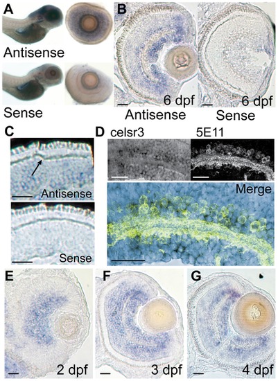
celsr3 is abundant within amacrine and ganglion cell layers of the retina. In situ hybridization was performed on WT animals using probes for exon1 of celsr3. A) Whole mounts of 6 dpf fish show localization to the brain and eye. When eyes were removed they show localization in the INL and ganglion cell layer. B and C) Cryosections of a 6 dpf eye show staining in the INL with abundance in the amacrine and ganglion cell layers. Sense controls show no staining. Arrow in C points to unlabeled horizontal cells. D) Slides were probed for celsr3 message and then probed with the anti-amacrine antibody 5E11 confirming presence of the message within amacrine cells. E–G) A time series of celsr3 message localization shows accumulation around the IPL at all ages from 2–4 dpf. Scale bars are 20 μm. |
| Gene: | |
|---|---|
| Antibody: | |
| Fish: | |
| Anatomical Terms: | |
| Stage Range: | Long-pec to Day 6 |