
Fig. 4
- ID
- ZDB-IMAGE-110909-24
- Genes
- Antibodies
- Publication
- Lewis et al., 2011 - Celsr3 Is Required for Normal Development of GABA Circuits in the Inner Retina
- All Figures
- Figures for Lewis et al., 2011
|
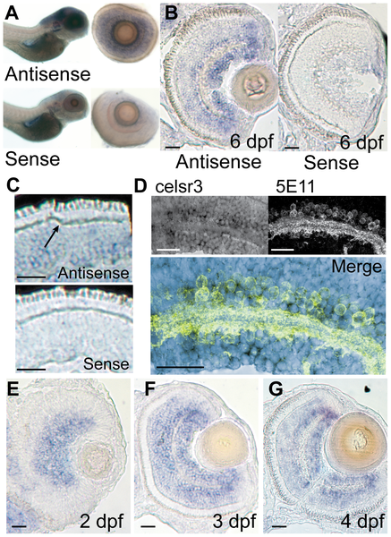
Fig. 4
celsr3 is abundant within amacrine and ganglion cell layers of the retina.
In situ hybridization was performed on WT animals using probes for exon1 of celsr3. A) Whole mounts of 6 dpf fish show localization to the brain and eye. When eyes were removed they show localization in the INL and ganglion cell layer. B and C) Cryosections of a 6 dpf eye show staining in the INL with abundance in the amacrine and ganglion cell layers. Sense controls show no staining. Arrow in C points to unlabeled horizontal cells. D) Slides were probed for celsr3 message and then probed with the anti-amacrine antibody 5E11 confirming presence of the message within amacrine cells. E–G) A time series of celsr3 message localization shows accumulation around the IPL at all ages from 2–4 dpf. Scale bars are 20 μm.