
Fig. 6
- ID
- ZDB-FIG-110811-44
- Publication
- Kikuchi et al., 2011 - tcf21+ epicardial cells adopt non-myocardial fates during zebrafish heart development and regeneration
- Other Figures
- All Figure Page
- Back to All Figure Page
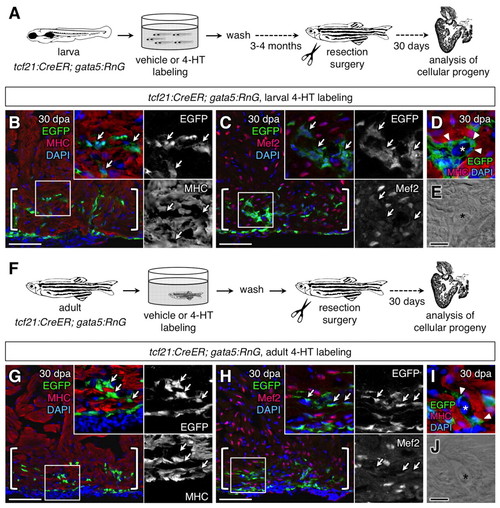
Epicardial cells contribute vascular support cells during heart regeneration. (A) Schematic of the experimental design. (B,C) Lineage-labeled EGFP+ cells (arrows, green) did not colocalize with Myosin heavy chain (MHC; B, red) or Mef2 (C, red) in 30 days post-amputation (dpa) regenerates of the larvally labeled animals (brackets; n=20). (D,E) EGFP+ cells (arrowheads) surrounds a vessel within the larvally labeled regenerate. Asterisk marks vascular lumen. (F) Schematic of the experimental design. (G,H) Lineage-labeled EGFP+ cells (arrows) did not colocalize with MHC (G) or Mef2 (H) in 30 dpa regenerates of the adult-labeled animals (brackets; n=6). (I,J) EGFP+ cells (arrowheads) surround a vessel within the adult-labeled regenerate. Asterisk marks vascular lumen. An antibody was used to detect EGFP in these experiments. Scale bars: 50 μm for B,C,G,H; 10 μm for D,E,I,J. |