
Fig. 2
- ID
- ZDB-FIG-110811-40
- Publication
- Kikuchi et al., 2011 - tcf21+ epicardial cells adopt non-myocardial fates during zebrafish heart development and regeneration
- Other Figures
- All Figure Page
- Back to All Figure Page
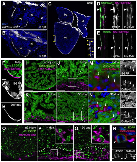
Zebrafish tcf21 regulatory elements drive epicardial-specific expression. (A-C) Cardiac tcf21:DsRed2 expression (white) at 3 dpf (A), 6 dpf (B) and adult (C) stages. Asterisks indicate extracardiac expression in A and B. Inset in C shows enlargement of area in yellow box. (D,E) tcf21:DsRed2+ epicardial cells (magenta) within the adult ventricular wall, assessed for wt1b:EGFP (D) or antibody staining for Raldh2 (E; green) colocalization. Arrowheads and arrows in C-E indicate the outer (epithelial) epicardial cell layer and EPDCs, respectively. (F-I) tcf21:DsRed2 (magenta) and cmlc2:EGFP (green) expression in distinct cells of 6 dpf (F-H) and adult (I) ventricles. An antibody was used to detect DsRed in F-H; this antibody also failed to detect DsRed2+EGFP+ cells in adult tcf21:DsRed2; cmlc2:EGFP ventricles (data not shown). (J-N) tcf21:DsRed2 (magenta or red) and cmlc2:EGFP (green) expression also mark distinct cells during regeneration. Arrows indicate tcf21:DsRed2+ nuclei in M and N. (O-R) tcf21:DsRed2 (magenta or red) and fli1:EGFP (green) do not colocalize in uninjured or regenerating adult ventricles. Insets in P and Q are enlarged views of boxed areas. Dashed lines indicate approximate amputation planes. at, atrium; ba, bulbus arteriosus; ve, ventricle. Scale bars: 100 μm for A-C; 10 μm for D-H,R; 50 μm for I-Q. |