
Fig. 3
- ID
- ZDB-FIG-110811-41
- Publication
- Kikuchi et al., 2011 - tcf21+ epicardial cells adopt non-myocardial fates during zebrafish heart development and regeneration
- Other Figures
- All Figure Page
- Back to All Figure Page
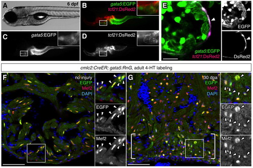
gata5 and tcf21 regulatory sequences drive expression that overlaps in the zebrafish epicardium. (A-E) A gata5:EGFP; tcf21:DsRed2 larva at 6 dpf. Insets are magnified images of the areas in white boxes in B-D. A single confocal slice visualizing gata5:EGFP; tcf21:DsRed2 ventricles shows colocalization limited to the epicardium (E, arrowhead). Each channel of the image is shown on the right (E). (F,G) The gata5:RnG indicator line efficiently reports induced Cre-mediated recombination in cardiomyocytes. cmlc2:CreER; gata5:RnG adults treated with 4-HT, showing EGFP labeling in Mef2+ cardiomyocytes nuclei of uninjured (F) and regenerated (G, brackets) cardiac tissue. Arrowheads in insets indicate EGFP immunofluorescence. Scale bars: 10 μm for E; 50 μm for F,G. |