|
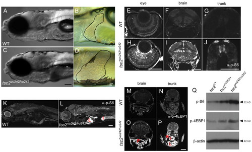
Upregulation of TORC1 activity in various tissues of tsc2vu242/vu242 mutants. (A–D) Homozygous mutant zebrafish have a deflated swim bladder and markedly enlarged liver at 7 dpf. Livers are outlined (B,D); asterisk indicates swim bladder. (E–P) Sections obtained from wild-type (WT) and tsc2vu242/vu242 embryos at 7 dpf and stained with anti-phospho-S6 ribosomal protein (Ser235/236) antibody (E–L) or anti-phospho-4E-BP1 (Thr37/46) (M–P). Phospho-S6 staining in the eye (E,H), brain (F,I) and trunk (G,J). Phospho-4E-BP1 staining in the brain (M,O) and trunk (N,P). (Q) Western blot using anti-phospho-S6 ribosomal protein antibody and anti-phospho-4E-BP1 of total protein lysates from 7-dpf wild-type, tsc2vu242/+ and tsc2vu242/vu242 larvae. Antibody staining for β-actin was used as a loading control. P, pharynx; K, kidney; I, intestine; L, liver. Scale bars: 100 μm.
|