|
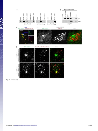
Rab11-GTP binds to Rabin8 as an effector through the Rabin8 COOH-terminal domain, colocalizes with Rabin8 in the endosome recycling compartment (ERC) before ciliogenesis, and is associated with the ciliogenesis and BBS pathway (related to Fig. 3). (A) Rab11aGTP selectively associates with the Rabin8 COOH-terminal membrane-targeting domain. Rab constitutively active (Q-L) mutants and the GDP-bound dominant negative Rab (T- or S-N) mutants were tested by yeast two-hybrid analysis against Rabin8 wild type and truncation mutants. (B) Effector binding of Rabin8 to Rab11 in mammalian cells. Equal amounts of control and nucleotide-exchanged IMCD3 LAP(S-protein-TEV-GFP-tag)-Rabin8 lysate were mmunoprecipitated with anti-GFP antibodies and immunoblotted with GFP and pan-Rab11 antibodies. (C) Rabin8 and Rab11a colocalize on vesicles near the cilia base. IMCD3 LAP-Rabin8 cells transiently expressing tRFP-Rab11a Q70L for 48 h were stained with anti-Actub antibodies. Projected deconvolved cell stacks are shown. (Scale bar: 5 μm.) (D) Vesicular GFPRabin8 does not localize to the Golgi. Micrographs show RPE GFP-Rabin8 cells serum starved for 1 h, fixed with cold methanol, and stained with anti-GM130 antibodies. Images were captured and processed as described in Fig. S1B. (E) GFP-Rabin8 colocalizes with transferrin in the ERC. Micrographs show RPE GFPRabin8 cells serum starved for 1 h at 37 °C followed by incubation with Alexa Fluor 594 transferrin and imaged live by confocal laser scanning microscopy. (F) Rab11 isoform siRNA depletion. Immunoblots of RPE lysate 72 h after siRNA transfection. ON-TARGETplus oligonucleotides are for Rab11a 52GCAACAAUGUGGUUCCUAUUU and Rab11b 52GGGACGACGAGUACGACUAUU. (G) Rab11 is required for Rab8-dependent ciliary membrane formation. GFP-positive cilia structures were counted (as described in Fig. S1E) in Rab11 siRNA-treated RPE GFP-Rab8a cells (n = 75) after 6-h serum starvation. *P < 0.0001; **P < 0.04. Representative results from three independent experiments are shown. (H) Rab11 depletion by siRNA impairs ciliation in RPE cells. Actub-positive cilia were counted in control and Rab11a+Rab11b siRNA-depleted parental RPE cells (n = 75 cells; *P < 0.001). Representative data from three independent experiments are shown. (I) Rabin8 functions in melanosome retraction in zebrafish. Control 5-d embryos retract their melanosomes in an average time of 96 s (n = 25) after epinephrine treatment, whereas embryos injected with 10 ng Rabin8 MO retracted melanosomes significantly more slowly, in an average time of 148 s (n = 452; *P < 0.01). Error bars indicate SEM. (J) Expression of Rab11 isoforms in zebrafish. Rab11a (NM_001007359), Rab11b (NM_001002555), and a third isoform Rab11a-like (NM_200123) during zebrafish development was evaluated by RT-PCR. dfp, d postfertilization; hfp, h postfertilization. Rab11a is expressed during Kupffer vesicle (KV) development in zebrafish. (K) Rab11a dominant negative expression causes abnormalities in KV formation. Injection of Rab11a S25N mRNA (300 pg) caused KV defects in 70% of embryos compared with 6% of uninjected control embryos (*P < 0.0001). Injection with Rab11a Q70L did not significantly affect KV formation. (L) Rab11 functions in melanosome retraction in zebrafish. (Left) Representative images of melanosome localization in melanocytes from the head region of 5 dpf embryos after epinephrine treatment. (Scale bars: 100 μm.) (Right) Control embryos retract their melanosomes in an average time of 92 s (n = 16), whereas embryos injected with 2.5 ng Rab11a MO retracted melanosomes significantly more slowly, in an average time of 151 s (n = 42; P < 0.01). Coinjection with hRab11a mRNA restored normal melanosome retraction (93 s; n = 22). Error bars indicate SEM.

|
