
Fig. 1
- ID
- ZDB-FIG-110321-19
- Publication
- Wu et al., 2011 - SNW1 Is a Critical Regulator of Spatial BMP Activity, Neural Plate Border Formation, and Neural Crest Specification in Vertebrate Embryos
- Other Figures
- All Figure Page
- Back to All Figure Page
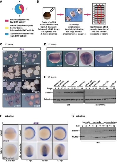
SNW1 was identified in a functional screen for neural crest fate in X. laevis. (A) A diagram of a stage 14 embryo depicting the classical model, which proposes that different levels of BMP signaling are required for ectodermal patterning and neural crest formation. (B) An overview of the functional screen. (C) The screen identified proteins whose overexpression modulated levels of Slug expression. 200 pg of mRNA expressing the X. tropicalis c-Myc, Dkk-1, Noggin2, or SNW1 was injected at the one-cell stage, with 250 pg of GFP mRNA as tracer. Control embryos received the GFP mRNA only. Embryos were fixed at stage 14 and analyzed for Slug expression by WISH. The number of embryos out of the total analyzed that showed the presented staining pattern is given. (D) X. laevis embryos were analyzed for SNW1 expression by WISH at the indicated stages. (E) Whole X. laevis embryo extracts were prepared from either wild-type (WT) embryos or embryos injected with 20 ng of a translation-blocking MO, MoSNW1a, at the one-cell stage, and analyzed by Western blotting using antibodies against SNW1 and Tubulin, the latter as a loading control. The developmental stages are indicated. (F) Zebrafish embryos were analyzed for SNW1 expression at the indicated times. Two gastrulation stages (the shield stage and 75% epiboly) and three segmentation stages (11, 12, and 13 hpf) are shown. Lateral views are shown, and for the later stages, also a dorsal-anterior view. (G) Whole zebrafish embryo extracts were prepared from wild-type embryos at the indicated times post-fertilization and analyzed by Western blotting using antibodies against SNW1 and MCM6, the latter as a loading control. The developmental stages are indicated. E, unfertilized egg. |
| Gene: | |
|---|---|
| Antibody: | |
| Fish: | |
| Anatomical Terms: | |
| Stage Range: | 1-cell to 14-19 somites |