|
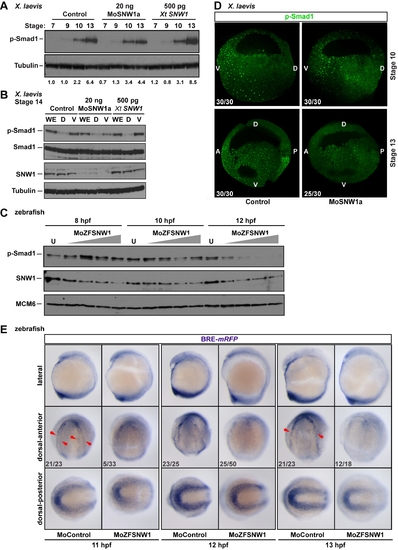
SNW1 regulates BMP activity in Xenopus and zebrafish embryos and is essential for BMP activity at the border between neural and non-neural tissue. (A) One-cell Xenopus embryos were either uninjected (control) or injected with 20 ng of MoSNW1a or 500 pg of X. tropicalis (Xt) SNW1. Embryos were harvested at the stages indicated, and whole embryo extracts were analyzed by Western blotting using antibodies against p-Smad1 and Tubulin, the latter as a loading control. Quantification of p-Smad1 levels relative to Tubulin is shown below the blots. (B) Xenopus embryos were either uninjected (control) or injected with 20 ng of MoSNW1a or 500 pg of X. tropicalis SNW1 mRNA at the one-cell stage, and bisected at stage 14 into dorsal (D) and ventral (V) halves. Whole cell extracts were analyzed by Western blotting using antibodies against SNW1, p-Smad1, Smad1, and Tubulin, the last as a loading control. WE, whole embryo. (C) Zebrafish embryos were either uninjected (U) or injected with increasing amounts (5, 10, 15, or 20 ng) of the splice-blocking MO MoZFSNW1. Embryos were harvested at 8, 10, or 12 hpf. Whole embryo extracts were analyzed by Western blotting using antibodies against SNW1, p-Smad1, and MCM6, the last as a loading control. (D) Knockdown of SNW1 strongly reduces ventral p-Smad1 levels at stage 13, but has no effect on the p-Smad1 ventral/dorsal gradient at stage 10. Embryos that were either uninjected (control) or injected with 20 ng of MoSNW1a at the one-cell stage were fixed at either stage 10 or stage 13, sagittally bisected along the midline, and immunostained with an antibody against p-Smad1. For these panels and subsequent p-Smad1 immunostaining, the specific p-Smad1 staining is nuclear and punctate. A, anterior; D, dorsal; P, posterior; V, ventral. (E) Transgenic BRE-mRFP embryos were injected with 15 ng of control MO or MoZFSNW1. They were fixed at 11, 12, and 13 hpf, when WISH was performed for mRFP, which indicates domains of BMP activity. In each case three different views of the same embryo are shown. In control embryos, mRFP is expressed in the tailbud (see dorsal-posterior view) and in a horseshoe-shaped domain at the dorsal anterior at 11 hpf (red arrowheads), which becomes increasingly sharpened at 12 and 13 hpf. In SNW1 morphants, the mRFP in this dorsal-anterior domain is reduced or absent. However, mRFP expression, and hence BMP activity, in the posterior is only slightly reduced or unchanged. In (D) and (E) the number of embryos out of the total analyzed that showed the presented staining pattern is given. In (E), the MO starts to knockdown SNW1 only from 8 hpf, and thus its effects become apparent only after 10 hpf. This explains why only a small percentage of embryos at 11 hpf shows the presented phenotype. By 13 hpf, when the MO effectively knocks down SNW1, 66% of the embryos show the phenotype.
|