
Fig. 8
- ID
- ZDB-FIG-110318-35
- Publication
- Felber et al., 2011 - Hedgehog signalling is required for perichondral osteoblast differentiation in zebrafish
- Other Figures
- All Figure Page
- Back to All Figure Page
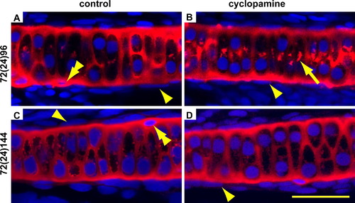
Blocking Hedgehog signalling does not lead to ectopic differentiation of chondrocytes in the perichondrium of the ceratohyal. Embryos were treated from 72 to 96 hpf then fixed immediately (A and B) or allowed to recover until 144 hpf (C and D). The cartilage is visualised with an antibody for Col II in red, and counterstained for nuclei with DAPI in blue. The arrowheads point to the flattened nuclei of the perichondrium which is present after EtOH (A and C) as well as cyclopamine treatment (B and D). There is no ectopic cartilage formation after cyclopamine treatment (B and D). However, Collagen 2 secretion is temporarily disrupted as shown by increased intracellular staining (arrow in B). There are unidentified cells on the edge of the cartilage that are embedded in Col II, but have not intercalated with the other chondrocytes and have highly condensed nuclei (double arrowheads in A and C). There are several of these cells on each ceratohyal and their presence was not affected by treatment. Please see Fig. 2 for abbreviations and notes. |
Reprinted from Mechanisms of Development, 128(1-2), Felber, K., Croucher, P., and Roehl, H.H., Hedgehog signalling is required for perichondral osteoblast differentiation in zebrafish, 141-152, Copyright (2011) with permission from Elsevier. Full text @ Mech. Dev.