
Fig. 2
- ID
- ZDB-FIG-110318-29
- Publication
- Felber et al., 2011 - Hedgehog signalling is required for perichondral osteoblast differentiation in zebrafish
- Other Figures
- All Figure Page
- Back to All Figure Page
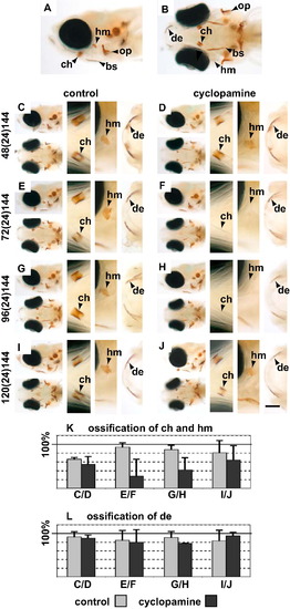
von Kossa staining of 6 dpf old zebrafish embryos shows the specific loss of chondral ossification after inhibition of the Hedgehog pathway. (A and B) Side and ventral views of the head indicating the positions of the bones analyzed in this study. (C, E, G and I) are control fish which were treated with EtOH. (D, F, H and J) are embryos treated with cyclopamine. The cyclopamine treatments from 72 to 96 hpf (E and F) and 96 to 120 hpf (G and H) show loss of chondral ossification of the ceratohyal and hyomandibula. Achondral ossification (the dentary and other bones) is unaffected. Embryos treated from 48 to 72 hpf (C and D) and 120 to 144 hpf (I and J) do not show a strong change in ossification. (K) shown the quantification of ossification of the ceratohyal and hyomandibula from the experiments shown in (C–J). (L) shown the quantification of ossification of the dentary from the experiments shown in (C–J). Notes on figures: Abbreviations: mx = maxilla; de = dentary; ch = ceratohyal and hm = hyomandibula; op = opercle; bs = branchiostegal ray. The timings of experiments shown in [Fig. 2, [Fig. 3], [Fig. 4], [Fig. 5], [Fig. 6] and [Fig. 7] are labeled with the following nomenclature: start of treatment (duration of treatment) time of fixation. Scale bars for high-magnification pictures = 50 μm. In [Fig. 2], [Fig. 3], [Fig. 4], [Fig. 5] and [Fig. 6], each panel shows low magnification side and ventral views to the left and high magnification ventral views to the right. Anterior is to the left. |
Reprinted from Mechanisms of Development, 128(1-2), Felber, K., Croucher, P., and Roehl, H.H., Hedgehog signalling is required for perichondral osteoblast differentiation in zebrafish, 141-152, Copyright (2011) with permission from Elsevier. Full text @ Mech. Dev.