
FIGURE
Fig. 3
- ID
- ZDB-FIG-110318-30
- Publication
- Felber et al., 2011 - Hedgehog signalling is required for perichondral osteoblast differentiation in zebrafish
- Other Figures
- All Figure Page
- Back to All Figure Page
Fig. 3
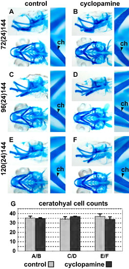
Alcian blue staining of 6 dpf old zebrafish embryos shows that chondrocyte differentiation and proliferation are unaffected by the loss of Hedgehog signalling after 72 hpf. (A, C and E) show the EtOH treated control fish. (B, D and F) show embryos treated with cyclopamine. There in no change in patterning or differentiation of the cartilage skeleton at the time points investigated as judged by morphology. (G) shown the quantification of the number of chondrocytes in the ceratohyal from the experiments shown in (A–F). Please see Fig. 2 for abbreviations and notes. |
Expression Data
Expression Detail
Antibody Labeling
Phenotype Data
Phenotype Detail
Acknowledgments
This image is the copyrighted work of the attributed author or publisher, and
ZFIN has permission only to display this image to its users.
Additional permissions should be obtained from the applicable author or publisher of the image.
Reprinted from Mechanisms of Development, 128(1-2), Felber, K., Croucher, P., and Roehl, H.H., Hedgehog signalling is required for perichondral osteoblast differentiation in zebrafish, 141-152, Copyright (2011) with permission from Elsevier. Full text @ Mech. Dev.