
Fig. 5
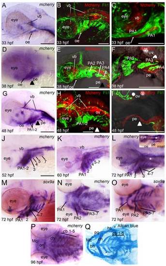
Reporter expression in Tg(Gli-d:mCherry) craniofacial region during embryogenesis and larval stages. Lateral (A–M) or ventral (N–Q) views, anterior to the left, of 33 hpf (A–C), 38 hpf (D–F), 48 hpf (G–I), 52 hpf (J), 60 hpf (K), 72 hpf (L–O) or 4.5 dpf (P,Q) transgenic fish stained with mCherry riboprobe (A,D,G,J,K,L,N,P) or sox9a riboprobe (M,O) for gene expression or Alcian blue (Q) to visualize cartilage elements. Confocal stack projections of Tg(Gli-d:mCherry) and Tg(Fli1:gfp) double transgenic fish (B,C,E,F,H,I). In these fish, reporter (red) and neural crest cells (green) can be visualized simultanenously. Of note, in (H–I) the reporter-positive pe tissue separated slightly from PA tissue during mounting of embryo. In (I) green-positive neural crest cells are not in the focal plane, despite residing just dorsal to mCherry expressing pe cells. (A–C) At 33 hpf, transgene is expressed in ventral brain and oral ectoderm. (D–F) At 38 hpf, expression persists in brain and ectoderm and expands to pharyngeal endoderm. (G–I) At 48 hpf, expression persists in brain and facial epithelium and expands to mesenchyme in anterior arches. (J–K) From 52–60 hpf, transgene expression expands to mesenchyme in all arches. (L–O) At 72 hpf, transgene expression in PA closely matches the mesenchymal neural crest marker sox9a. (P,Q) At 4.5 dpf, transgene expression persists in each arch and the pattern matches cartilage elements. Abbreviations: bh, basihyal; cb, ceratobranchial; ch, ceratohyal; hs, hyosymplectic; Mc, Meckel′s cartilage; oe, oral ectoderm; on, optic nerve; PA, pharyngeal arch; pe, pharyngeal endoderm; pq, palatoquadrate; tr, trabeculae; vb, ventral brain. |
| Genes: | |
|---|---|
| Fish: | |
| Anatomical Terms: | |
| Stage Range: | Prim-15 to Day 4 |