
Fig. 7
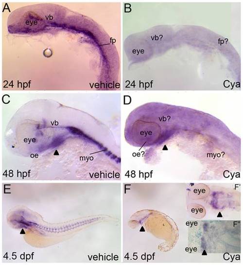
Reporter expression in Tg(Gli-d:mCherry) fish following severe reductions to Hh-signaling. Lateral (A–F) or ventral (F′,F") views, anterior to the right, of Tg(Gli-d:mCherry) fish treated with vehicle (A,C,E) or Cya (B,D,F,F′,F") and stained with mCherry riboprobe (A–F′) or dually stained with mcherry and Alcian blue (F"). Vehicle or Cya treatments began at dome stage and commenced just prior to sacrificing the embryos or larvae for staining analysis. (A,B) At 24 hpf, Cya treatments suppressed all head and trunk reporter expression in transgenic embryos. (C,D) At 48 hpf, Cya treatments reduced all reporter expression in transgenic fish, with the exception of a small expressing domain in the ventral head (arrowhead). (E,F) Reporter expression was virtually absent in Cya treated 4.5 dpf transgenic larvae, with the exception of the same mCherry expressing domain in the ventral head that was seen in 48 hpf aged embryos. (F′, F") Closer examination of the ventral head suggested that the mCherry expressing cells are within second arch mesenchyme and that no cartilage forms in this arch or any other PA in Cya treated larvae. Abbreviations: fp, floor plate; myo, myotome; vb, ventral brain. |