
Fig. S4
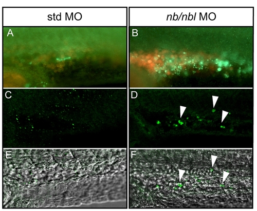
Caspase-3 activation in nb/nbl morphants at 28–30 hpf. A–B. Whole-mount immunofluorescence to detect caspase-3 activation (green signal), detailed view of the ICM region of 26–28 hpf Tg(gata1:dsRed) embryos injected with std MO (A) and nb/nbl MO (B). C–F. Single optical sections of 26–28 hpf control and nb/nbl MO-injected embryos in which whole-mount immunofluorescence for caspase-3 activation (green signal) was performed. Fluorescent images (C, D) were merged with bright field images (E, F). Detailed view of the ICM region. Caspase-3 activation can be detected in erythroid cells of nb/nbl morphants (white arrowheads; D, F). |