
Fig. 8
- ID
- ZDB-FIG-101208-29
- Publication
- Tu et al., 2010 - Clonal analyses reveal roles of organ founding stem cells, melanocyte stem cells and melanoblasts in establishment, growth and regeneration of the adult zebrafish fin
- Other Figures
- All Figure Page
- Back to All Figure Page
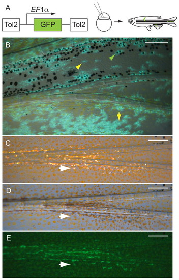
Melanocytes and xanthophores come from the same founding stem cells (mFSCs), and iridophores arise from a distinct founding stem cell (iFSC). (A) EF1α>GFP lineage marker transposon was injected into one- or two-cell stage mlpha embryos. The mosaic embryos were reared to maturity, then screened for GFP+ clones in the fins. (B) Fluorescence image of a labeled caudal fin clone in which both the melanocytes and xanthophores are labeled with GFP. Green arrowhead shows a labeled melanocyte; yellow arrowhead shows a covert xanthophore (lightly pigmented xanthophores within the melanocyte stripe) (Hirata et al., 2003; Hirata et al., 2005) and yellow arrow shows an overt xanthophore. (C-E) An iridophore clone. White arrows indicate the same iridophore in the three light conditions: (C) incident light, (D) transillumination and (E) GFP fluorescence. Scale bars: 0.2 mm. |