
Fig. 4
- ID
- ZDB-FIG-101208-25
- Publication
- Tu et al., 2010 - Clonal analyses reveal roles of organ founding stem cells, melanocyte stem cells and melanoblasts in establishment, growth and regeneration of the adult zebrafish fin
- Other Figures
- All Figure Page
- Back to All Figure Page
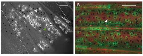
Melanocytes descended from different mFSCs intermix. (A) Fluorescence image of labeled anal fin from a mosaic animal generated by injection of fTyrp>GFP transposon. Non-labeled melanocytes (white arrowhead) are present within the territory of the GFP+ melanocytes (green arrowhead). Broken lines show the border of the clone. (B) Fluorescence image of central stripe from a fish co-injected with fTyrp>GFP and fTyrp>mCherry transposons. All melanocytes in this area are labeled either with GFP (green) or mCherry (red). Arrowhead indicates what appears to be a melanocyte of both colors, but in fact is two overlapping red and green melanocytes at different focal depth in the fin. Scale bars: 0.2 mm. |