
FIGURE
Fig. 2
- ID
- ZDB-FIG-101208-23
- Publication
- Tu et al., 2010 - Clonal analyses reveal roles of organ founding stem cells, melanocyte stem cells and melanoblasts in establishment, growth and regeneration of the adult zebrafish fin
- Other Figures
- All Figure Page
- Back to All Figure Page
Fig. 2
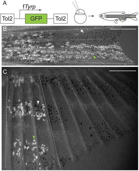
Labeled melanocyte clones extend proximo-distally in the caudal and anal fins. (A) fTyrp>GFP lineage transposon (Zou et al., 2006) was injected into one- or two-cell embryos, which were reared to maturity prior to screening for melanocyte clones in fins. (B,C) Examples of labeled melanocyte clones in center stripe, of caudal fin (B), and anterior 4 fin rays, of anal fin (C). White arrowheads indicate GFP- melanocytes; green arrowheads indicate GFP+ melanocytes; broken white lines show outer boundaries of clones. Scale bars: 0.5 mm. |
Expression Data
Expression Detail
Antibody Labeling
Phenotype Data
Phenotype Detail
Acknowledgments
This image is the copyrighted work of the attributed author or publisher, and
ZFIN has permission only to display this image to its users.
Additional permissions should be obtained from the applicable author or publisher of the image.
Full text @ Development