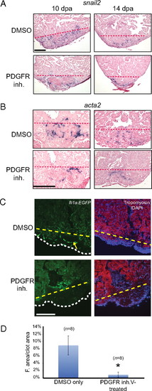
PDGF signaling is required for coronary blood vessel formation during zebrafish heart regeneration. (A) Treatment of wild-type fish with DMSO or PDGFR inhibitor (inh.) from 2–14 dpa. snail2 ISH was performed using 10- and 14-dpa hearts. (Scale bar = 100 μm.) (B) Treatment of wild-type fish with DMSO or PDGFR inhibitor from 2–14 dpa. acta2 ISH was performed using 10- and 14-dpa hearts. (Scale bar = 100 μm.) (C) Treatment of fli1a:EGFP fish with PDGFR inhibitor from 2–14 dpa. Hearts were collected and processed at 14 dpa (DAPI staining, blue; tropomyosin staining, red). An example of blood vessel formation (green) in the regenerating hearts is marked by the yellow arrow. The dashed lines mark the approximate position of the amputation plane in A–C. The white dashed line indicates the outline of the fibrin clot in the regenerating hearts. (Scale bar = 100 μm.) (D) Quantification of blood vessel formation. The total fluorescent (F.) area was measured relative to total clot area (n = 8 hearts for DMSO and PDGFR inhibitor treatment). *P < 0.0001.
|
