
Fig. S6
- ID
- ZDB-FIG-100903-53
- Publication
- Wang et al., 2010 - Moesin1 and Ve-cadherin are required in endothelial cells during in vivo tubulogenesis
- Other Figures
- All Figure Page
- Back to All Figure Page
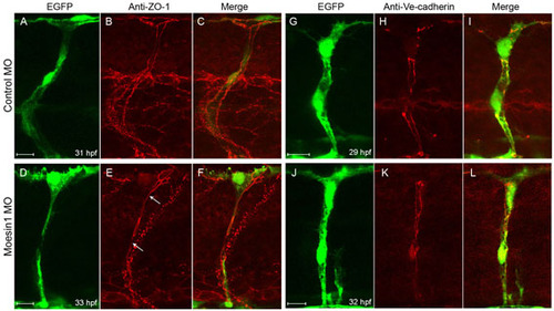
Knockdown of Moesin1 results in partial loss of adherens junctions without affecting ZO-1-associated junctions between endothelial cells in the ISVs. (A-F) Confocal images of the ISVs in Tg(fli1:egfp)y1 embryos that were probed for ZO-1 (red) at 34 hpf and 36 hpf. (A-C) In control MO-injected embryos, two ZO-1-associated junctions that run lengthwise along the ISV are detected. (D-F) In Moesin1 knockdown embryos, the ZO-1-associated tight junctions are observed along the length of the ISV (arrow), but the junctions are not separated from one another as in the control embryos. (G-L) Confocal images of the ISVs from Tg(fli1:egfp)y1 embryos are labeled with Ve-cadherin (red) at 29 hpf and 32 hpf. (G-I) Two strands of Ve-cadherin labeling that run lengthwise along the ISV are observed in control embryos. (j-l) Moesin1 knockdown causes partial loss of Ve-cadherin labeling (arrow), with junctions between cells appearing as beads (arrowhead) on a string rather than a continuous junction. (a-l) Lateral images, dorsal is up, anterior is to the left. Scale bars: 10 μm. |