
Fig. 3
- ID
- ZDB-FIG-100809-5
- Publication
- Schröter et al., 2010 - Segment Number and Axial Identity in a Segmentation Clock Period Mutant
- Other Figures
- All Figure Page
- Back to All Figure Page
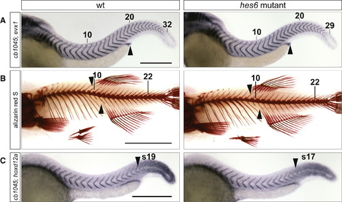
hes6 Mutants Have a Reduced Number of Embryonic and Adult Segments, and the Change in Segment Number Is Distributed across Axial Regions (A) Embryos at 48 hpf were stained with the myotome boundary marker cb1045 and evx1, which labels the proctodeum (arrowhead). hes6 mutants have fewer myotomes than their wild-type siblings, and their proctodeum aligns with the 16th segment instead of the 17th or 18th as in the wild-type. The tenth, 20th, and last segment are indicated. The scale bar represents 0.3 mm. (B) Skeletal stains of 2-month-old wild-type and hes6 mutant fish. The mutant has fewer vertebrae and ribs but an otherwise normal morphology of the vertebral column. The anterior insertion sites of the anal and dorsal fin (arrowheads) align with a higher vertebral number in wild-type compared to hes6 mutant fish. The 10th and 22nd vertebrae are indicated. The scale bar represents 0.5 cm. (C) Embryos at 25 hpf were costained for cb1045 and hoxd12a expression. The anterior border of hoxd12a expression (arrowhead) coincides with a lower segmental count in hes6 mutant compared to wild-type embryos. The scale bar represents 0.3 mm. See also Figure S2, Table S2, and Table S3. |
| Genes: | |
|---|---|
| Fish: | |
| Anatomical Terms: | |
| Stage Range: | Prim-5 to Long-pec |
| Fish: | |
|---|---|
| Observed In: | |
| Stage Range: | Prim-5 to Days 45-89 |