
FIGURE
Fig. S4
Fig. S4
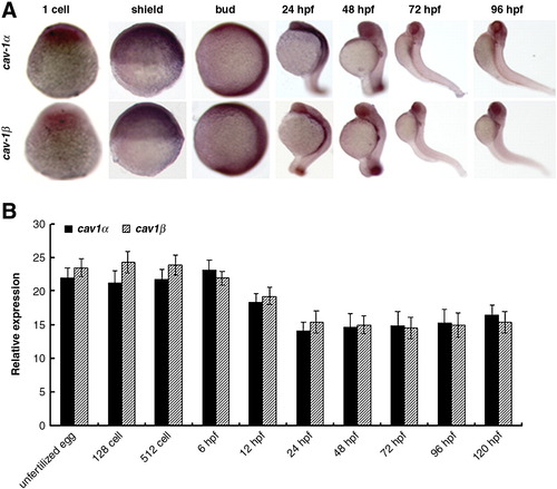
Spatiotemporal expression of cav-1 during embryogenesis. (A) Whole-mount in situ hybridization was performed with antisense RNA probes in embryos at indicated stages. 1-cell and shield stages, lateral view with the animal pole to the top; Bud stage to 96 hpf, lateral view with the anterior to the top. (B) Relative expression of Cav-1α or -1β during embryogenesis. Embryos at indicated stages were collected for total RNA isolation and subjected to real-time PCR analysis. Expression of 18s ribosomal RNA was used as an internal control. Values are given as mean ± standard deviation, n = 3. |
Expression Data
Expression Detail
Antibody Labeling
Phenotype Data
Phenotype Detail
Acknowledgments
This image is the copyrighted work of the attributed author or publisher, and
ZFIN has permission only to display this image to its users.
Additional permissions should be obtained from the applicable author or publisher of the image.
Reprinted from Developmental Biology, 344(1), Mo, S., Wang, L., Li, Q., Li, J., Li, Y., Thannickal, V.J., and Cui. Z., Caveolin-1 regulates dorsoventral patterning through direct interaction with beta-catenin in zebrafish, 210-223, Copyright (2010) with permission from Elsevier. Full text @ Dev. Biol.