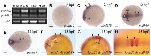
Pcdh19 expression in 6-13 hpf zebrafish embryos. (A) RT-PCR analysis of Pcdh19 isoforms 1 and 2 expression in embryonic zebrafish using total RNAs. RT-PCR for cdh1 was performed as loading control. The remaining panels show whole mount embryos labeled with Pcdh19 cRNA probes (B-E), Pcdh19 and pax2a cRNA probes (F), or Pcdh19 and krox20 cRNA probes (G,H). (B,C) Lateral views of the entire embryos (head towards the lower left corner for C). (D,E) Dorsal views (anterior to the left) of the entire embryos. (F,G) Lateral views of the anterior half of the embryos (anterior to the left and dorsal up), while (H) is a dorsal view of the presumptive hindbrain region of an embryo (anterior to the left). The arrow and arrowhead in (C, D, F, G and H) point to the first and second Pcdh19 expression domains, respectively, in the presumptive hindbrain. The two arrows in panel E indicate Pcdh19 expression in the middle neural keel. Abbreviations: bmh, boundary of the mid- and hindbrains; ep, eye premordium; f, presumptive forebrain; h, presumptive hindbrain; r3 and r5, rhombomeres 3 and 5, respectively. Scale bars, 100 μm.
|
