|
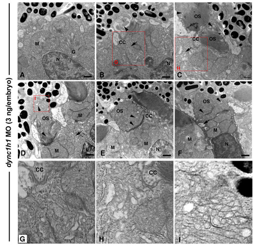
Dync1h1 knock-down causes dose-sensitive outer segment defects. TEM of retinas from 3-dpf embryos injected with a high dose (8 ng/embryo) of dync1h1 morpholino (MO). (A) A cone cell with Golgi apparatus (G) localized in the central ellipsoid region and mitochondria (M) located near the nucleus (N). (B-I) Cone cells with accumulation of vesicles (arrows) at the base of the connecting cilium (CC) and presence of disorganized disc structures within the outer segment (OS; arrowheads). (D-F) Severely disrupted outer segments showing vesiculation and tubulation of discs structures distally (arrowheads), yet normal discs at the base. Large vacuoles and vesicle accumulations were found at the base of the connecting cilium (arrows). The condensed cell in (F) is consistent with the morphology of early stage apoptosis. (G-I) Higher magnification insets of the red boxed regions. Scale bars: 0.5 μm in (A-F).
|

