
Fig. 4
- ID
- ZDB-FIG-100616-95
- Publication
- Insinna et al., 2010 - Analysis of a zebrafish dync1h1 mutant reveals multiple functions for cytoplasmic dynein 1 during retinal photoreceptor development
- Other Figures
- All Figure Page
- Back to All Figure Page
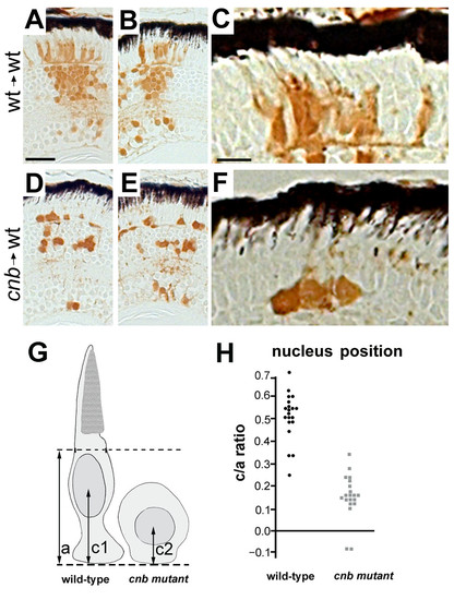
Photoreceptors from cnb/dync1h1 mutants show cell-autonomous defects in morphogenes. (A-F) Images of histology sections from genetically mosaic retinas of wild-type (wt) donor, wild-type host (A-C) or cnb donor, wild-type host (D-F). Two examples are shown at low magnification to indicate overall clone size and one example at higher magnification to show photoreceptor morphology. Donor cells are label with the brown lineage tracer. Black retinal pigment epithelium cells provide reference for orientation. Note the lack of elongated morphology in cnb photoreceptor cells. Scale bars: 40 μm in (A, B, D, E); 15 μm in (C, F). (G) Schematic showing how nuclear position was evaluated. Dashed lines represent the external limiting membrane (upper) and apical side of the outer plexiform layer (bottom) for the host retina. (H) Scatter plot of nuclear position (c/a ratio) for donor cells of wild-type (black, left side) or cnb (grey, right side) genotypes. |