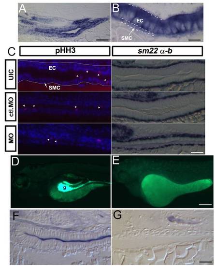
Expression of gata6 in the gut and characterization of differentiation defects in the miR-145 morphants. (A)Alongitudinal section of a 96 hpf wild-type embryo shows strong expression of gata6 in gut epithelial and smooth muscle cells. (B) Enlargement of (A) highlights staining of gata6 in both layers. White dashed lines distinguish the smooth muscle cell layer and epithelial cell layer. (C) Longitudinal sections of phosphohistone H3 (pHH3) staining (red) in the 96 hpf gut. Nuclei were counterstained with DAPI (blue). No significant difference of pHH3 positive cells (white arrowheads) was found in the different experimental groups in either epithelial or smooth muscle layers. The white line in the uninjected embryo highlights the gut epithelium. In the second column, longitudinal sections of sm22α-b in situ hybridization in 96 hpf embryos shows specific staining in the smooth muscle cell layer, and highlights abnormal morphology of both epithelial and smooth muscle cells in miR-145 morphants as compared to controls. (Dand E) The wild-type gut (g) is strongly fluorescent at 120 hpf after DAF-2DA staining (Green) in comparison with miR-145 morphants. (F and G) Strong alkaline phosphatase activity is observed in the gut lumen of wild-type embryos but is absent in miR-145 morphants. SMC, smooth muscle cell (white arrow); EC, epithelial cell; ctl. MO, control morpholino. (Scale bars, A: 50 μm; B: 10 μm; C and G; 25 μm; D: 200 μm.)
|
