
Fig. 7
- ID
- ZDB-FIG-100504-43
- Publication
- Grant et al., 2010 - The neuroepithelial basement membrane serves as a boundary and a substrate for neuron migration in the zebrafish hindbrain
- Other Figures
- All Figure Page
- Back to All Figure Page
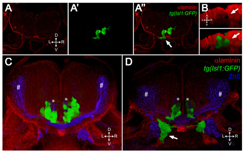
FBMNs exit the hindbrain through holes in the ventral Laminin-containing basement membrane that can be phenocopied by Laminin knockdown. (A-A″) Single XY section of a vibratome cross-section through a 48-hpf aPKCλ+ζ, tg(isl1:GFP) double morphant stained for Laminin (red). Ventrally mismigrating FBMNs exit the hindbrain through a hole in Laminin (arrow). (B) Ventral view of a three-dimensional reconstruction of the same coronal section showing the Laminin hole and the mismigrating FBMNs (arrows). (C, D) Three-dimensional reconstructions of 70 μm thick vibratome cross-sections through 48- hpf wild-type (C) and Lamininα 1 morpholino-injected tg(isl1:GFP) embryos (D) stained for Laminin (red) and Zn5 (blue) showing that Laminin knockdown results in ventrally mismigrated FBMNs (arrow) that exit the hindbrain while leaving abducens motor neurons (asterisks) and commissural interneurons (hash symbols) unaffected. ZN5-staining neurons outside the hindbrain are the sensory neurons of the acoustic nerve (nVIII). |