
Fig. 6
- ID
- ZDB-FIG-100504-42
- Publication
- Grant et al., 2010 - The neuroepithelial basement membrane serves as a boundary and a substrate for neuron migration in the zebrafish hindbrain
- Other Figures
- All Figure Page
- Back to All Figure Page
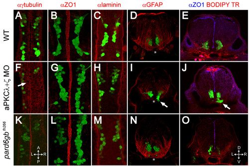
Removal of aPKC or Pard6gb results in defects of apical midline formation and maintenance. (A-C, F-H, K-M) Single XY sections (A, C, F, H, K, M) or three-dimensional reconstructions (B, G, L) of 24 hpf Isl1:GFP transgenic embryos stained for γ-tubulin (A), ZO1 (B), or Laminin (C) show that while apical tight junctions and basal basement membrane are formed correctly in the ventral hindbrain of embryos lacking aPKC or Pard6gb (B, C, G, H, L, M), there are subtle defects in neuroepithelial progenitor polarity reflected in misaligned centrosomes (F) (arrow). (D, I, N) Three-dimensional reconstructions of 70 μm thick 48-hpf cross-sections stained with α GFAP (α-Glial fibrillary acidic protein) show that while staining is reduced in aPKCλ+ζ double knockdown embryos, the absence of radial glial endfeet is unlikely to explain ventral mismigration as endfeet are absent from the most ventromedial region (where mismigration occurs in morphants, arrow) of both wild0type embryos and morphants (asterisks). (E, J, O) Single XY optical sections of 48-hpf vibratome cross-sections stained for ZO1 and with BODIPY TR methylester dye show reduced staining in aPKCλ+ζ double morphants in which ventral mismigration is occurring (J, arrow) or pard6gbfh266 mutants (O), indicating that apical tight junctions are lost between 24 and 48 hpf in these embryos. |