
Fig. 3
- ID
- ZDB-FIG-100504-39
- Publication
- Grant et al., 2010 - The neuroepithelial basement membrane serves as a boundary and a substrate for neuron migration in the zebrafish hindbrain
- Other Figures
- All Figure Page
- Back to All Figure Page
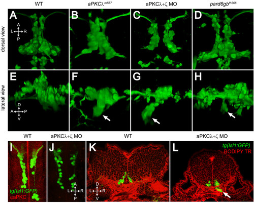
PAR-aPKC complex disruption causes a ventral mismigration in which FBMNs escape the hindbrain ventrally. (A-H) Three-dimensional reconstructions of confocal Z-stacks of the Isl1:GFP positive facial motor neurons at 48 hpf in wild-type (WT) (A, E), aPKCλm567 homozygous mutant (B, F), aPKCλ+ζ double morpholino knockdown (C, G), and pard6gbfh266 homozygous mutant embryos (D, H) shown in dorsal (A-D) and lateral (E-H) views. In aPKCλm567, aPKCλ+ζ double morpholino knockdown, and pard6gbfh266 embryos (F, G, H), a subset of FBMNs mismigrates ventrally (arrows) forming ectopic ventral clusters. (I, J) Maximum intensity projections of confocal Z-stacks of 24-hpf WT (I) and aPKCλ+ζ MO (J) embryos stained with an antibody that recognizes both aPKCs reveals that double morpholino knockdown reduces aPKC to undetectable levels. (K, L) Vibratome coronal sections through 48-hpf WT (K) and aPKCλ+ζ MO (L) embryos at 48 hpf counterstained with BODIPY TR methylester dye that stains all membranes reveals that morphant FBMNs are found outside the hindbrain (arrow). In addition, the cross-sectional area of the aPKCλ+ζ MO hindbrain is reduced and the ventricle has not inflated. |