
Fig. 7
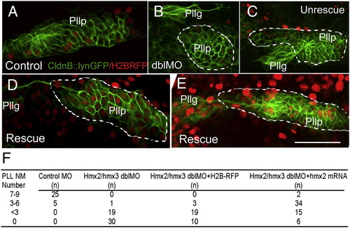
Mosaic expression of Hmx partially rescues the phenotype in posterior lateral line of hmx2/hmx3 morphants. Anterior to the left. The posterior lateral line primordium is labeled with the membrane GFP in CldnB::lynGFP transgenic embryo. (A) Normal control primordium injected with H2BRFP RNA (red) at 24 hpf. (B–E) Rescue experiment with co-injection of Hmx2 and H2BRFP RNAs (red) in hmx2/hmx3 morphant embryos. (B, C) Abnormal primordium of morphants without Hmx2 and H2BRFP injection. (D, E) Rescued primordium following Hmx2 and H2BRFP injection in morphants. (F) Summary of the number of neuromasts formed in each injection group. Embryos were stained for the endogenous alkaline phosphatase activity at 3dpf and number of neuromasts in each embryo counted. Sample size (n) is indicated. Scale bar: 50 μm. Abbreviations: Pllp, posterior lateral line primordium; PLL NM, posterior lateral line neuromast. |
| Gene: | |
|---|---|
| Fish: | |
| Knockdown Reagents: | |
| Anatomical Term: | |
| Stage: | Prim-5 |
Reprinted from Developmental Biology, 339(2), Feng, Y., and Xu, Q., Pivotal role of hmx2 and hmx3 in zebrafish inner ear and lateral line development, 507-518, Copyright (2010) with permission from Elsevier. Full text @ Dev. Biol.