|
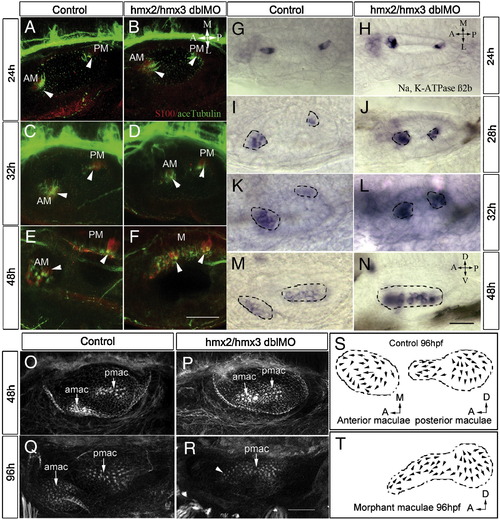
Reduced number of hair cells and progressive fusion of two maculae in hmx2/hmx3 morphants. Anterior to the left, dorsal view in panels A–L and lateral view in panels M–R. (A–F) Hair cells are labeled with antibodies for S-100 (red) and acetylated-tubulin (green) at the stages indicated showing progressive fusion of the anterior (utricular) maculae with the posterior (saccular) maculae at the medial wall in the morphants. (G–N) In situ hybridization with Na,K-ATPase β2b probe to reveal mature hair cells, showing a reduced number in the anterior maculae (amac) and fusion with the posterior maculae (pmac). (O–R) Rhodamine–phalloidin staining of the F-actin of the hair bundles in control (O, Q) and morphants (P, R). (S) Illustration of control amac and pmac from 9 ears. (T) Illustration of fused maculae in morphants from 8 ears. Arrowheads indicate hair cells. Scale bars: (A–F) 50 μm, (G–R) 25 μm. Abbreviations: AM, anterior maculae; PM, posterior maculae; M, maculae fusion.
|