|
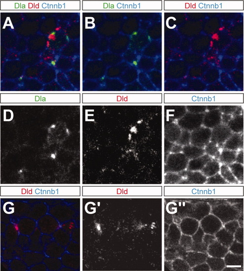
Dla and Dld protein subcellular localization. Lateral views of two different 26 hours postfertilization (hpf) embryos (A-F and G-G″). A: Triple staining of Dla antibody (green), Dld antibody (red), and Beta-catenin antibody (Ctnnb1, blue) showing that both Dla and Dld often localize to the cell cortex and/or membrane. B: Dla (green) colocalizes with Ctnnb1 (blue). C: In many cases, Dld (red) overlaps with Ctnnb1 (blue) staining. D-F show the green, red and blue channels, respectively. G: Double staining of Dld antibody (red) and Beta-catenin antibody (Ctnnb1, blue) showing that in the same region of the nervous system, Dld can be in the cell cortex and/or membrane (left side of panel) and also cytoplasmic (right side of panel). G′,G″ show the red and blue channels, respectively. Scale bar = 2.7 μm.
|