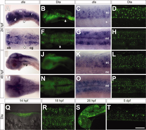
Comparison of dla mRNA and Dla protein expression patterns in the developing zebrafish embryo. A-D,I-L,S-T show lateral views, E-H,M-P,R show dorsal views. A,B: dla mRNA (A) and Dla protein (B) are expressed in a regionally restricted pattern in the developing brain at 24 hours postfertilization (hpf). C,D: Arrows mark the dla/Dla-free zone. Scattered dla mRNA (C) and Dla protein (D) distribution in the spinal cord. E: Defined dla mRNA expression in strings of cells in the presumptive brain. F: Dla protein appears more punctate than dla mRNA, but is expressed in the same brain regions. G,H: Spinal cord dla mRNA expression (G) compared with Dla protein labeling (H). I,J: At 48 hpf, dla mRNA (I) and Dla protein (J) distributions are more defined than at earlier stages. K,L: dla mRNA (K) and Dla protein (L) are widely expressed throughout the spinal cord. M: Dorsal view highlighting the highly ordered pattern of dla mRNA in the rhombomeres. N: Dla protein expression in the rhombomeres resembles dla mRNA distribution. O,P: dla mRNA (O) and Dla protein (P) labeling suggest a regionally conserved pattern in the spinal cord. Q: At 14 hpf, Dla protein is expressed throughout the neural plate, shown in a transverse view. R: Dla protein distribution at 18 hpf. S: Dla protein is localized in cell clusters in the developing brain at 26 hpf. T: At 5 dpf, Dla protein expression is maintained in some cells in the spinal cord. t, telencephalon; d, diencephalon; m, mesencephalon; r, rhombencephalon; sc, spinal cord; s, somite; ob, olfactory bulb; cg, cranial ganglia; trg, trigeminal ganglia; ml, midline; nc, notocord. Scale bars = 77 μm in A,B,E,F,I,J, 39 μm in C,D,G,H, 47 μm in M,N, 53 μm in K,L,O,P,Q, 33 μm in R, 65 μm in S, 25 μm in T.
|
