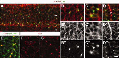
Dla protein subcellular localization on the cell cortex and/or membrane. Lateral (A-C) and transverse (D) views of 26 hours postfertilization (hpf) -old embryos labeled with Beta-catenin antibody (Ctnnb; red) and Dla antibody (green). A: Membrane localization of Dla, visible as dots or lines. B: Higher magnification reveals a putative cell to cell contact marked by Dla protein. (B′,B″) show the red and green channels respectively. C: Multiple Dla clusters suggest contact with several adjacent cells. C′,C″ show the red and green channels respectively. D: Transverse section showing the localization of Dla in the spinal cord relative to the midline. D′,D″ show the red and green channels respectively. E: Lateral view of 26-hpf-old embryo showing membrane green fluorescent protein (GFP; memGFP) [Tg(Bactin:HRAS-EGFP); green] and Dla antibody (red). F: Transverse section of adult brain labeled with Dla antibody showing distribution around entire cells. G: Lateral view of 26 hpf spinal cord labeled with Dla antibody showing punctate distribution. fp, floor plate; m, midline; sc, spinal cord. Scale bars = 5 μm in A,D, 3 μm in B,C, 4 μm in E-H″.
|
