
FIGURE
Fig. 49
- ID
- ZDB-FIG-091217-40
- Publication
- Parichy et al., 2009 - Normal table of postembryonic zebrafish development: Staging by externally visible anatomy of the living fish
- Other Figures
-
- Fig. 1
- Fig. 2
- Fig. 5
- Fig. 6
- Fig. 8
- Fig. 10
- Fig. 11
- Fig. 13
- Fig. 14
- Fig. 16
- Fig. 17
- Fig. 18
- Fig. 19
- Fig. 21
- Fig. 22
- Fig. 23
- Fig. 24
- Fig. 25
- Fig. 26
- Fig. 27
- Fig. 28
- Fig. 32
- Fig. 33
- Fig. 34
- Fig. 35
- Fig. 36
- Fig. 37
- Fig. 38
- Fig. 39
- Fig. 40
- Fig. 41
- Fig. 42
- Fig. 43
- Fig. 44
- Fig. 45
- Fig. 46
- Fig. 47
- Fig. 48
- Fig. 49
- Fig. 50
- Fig. 51
- Fig. 52
- Fig. 53
- Fig. 54
- Fig. 55
- Fig. 56
- Fig. 57
- All Figure Page
- Back to All Figure Page
Fig. 49
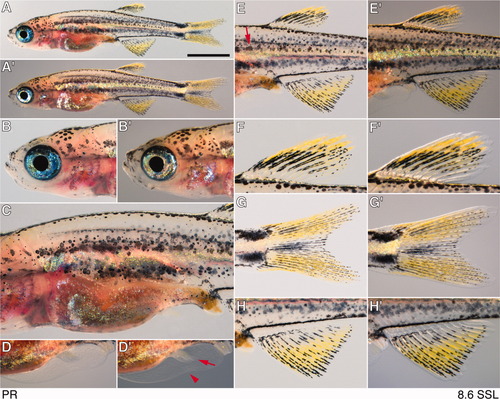
Pelvic fin ray appearance; PR, 8.5 mm SL (standard length). A,A′: Whole body. Scale bar = 2 mm. B,B′: Head. C: Anterior and middle trunk. D,D′: Pelvic fin with first rays (arrow) as well as minor lobe of fin fold and site of resorption (arrowhead). E,E′: Middle trunk showing dorsal and anal fins and pigment pattern. Residual embryonic/early larval melanophores occur near the horizontal myoseptum (arrow). F,F′: Dorsal fin. G,G′: Caudal fin, showing emergence of first stripes. H,H′: Anal fin. |
Expression Data
Expression Detail
Antibody Labeling
Phenotype Data
Phenotype Detail
Acknowledgments
This image is the copyrighted work of the attributed author or publisher, and
ZFIN has permission only to display this image to its users.
Additional permissions should be obtained from the applicable author or publisher of the image.
Full text @ Dev. Dyn.