
Fig. 27
- ID
- ZDB-FIG-091217-21
- Publication
- Parichy et al., 2009 - Normal table of postembryonic zebrafish development: Staging by externally visible anatomy of the living fish
- Other Figures
-
- Fig. 1
- Fig. 2
- Fig. 5
- Fig. 6
- Fig. 8
- Fig. 10
- Fig. 11
- Fig. 13
- Fig. 14
- Fig. 16
- Fig. 17
- Fig. 18
- Fig. 19
- Fig. 21
- Fig. 22
- Fig. 23
- Fig. 24
- Fig. 25
- Fig. 26
- Fig. 27
- Fig. 28
- Fig. 32
- Fig. 33
- Fig. 34
- Fig. 35
- Fig. 36
- Fig. 37
- Fig. 38
- Fig. 39
- Fig. 40
- Fig. 41
- Fig. 42
- Fig. 43
- Fig. 44
- Fig. 45
- Fig. 46
- Fig. 47
- Fig. 48
- Fig. 49
- Fig. 50
- Fig. 51
- Fig. 52
- Fig. 53
- Fig. 54
- Fig. 55
- Fig. 56
- Fig. 57
- All Figure Page
- Back to All Figure Page
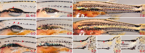
Development of iridophore pattern. Shown are multiple individuals (standard length [SL] at lower right). A: Development of iridophores in the nascent interstripe of the anterior trunk. 4.5, iridophores are first present only internally, covering the swim bladder (arrowhead). 5.0, iridophores immediately under the skin first arise near the swim bladder, over the ventral-lateral surfaces of a few myotomes. 5.7-7.3, the initial patch spreads more posteriorly (arrowheads indicate posteriormost reflective cells). B: A continuous iridophore interstripe is formed as cells in the anterior become contiguous with a patch at the base of the tail. C: Development of iridophore patch near the site of notochord flexion (anteriormost reflecting cells indicated by arrowheads). |