
Fig. 23
- ID
- ZDB-FIG-091217-17
- Publication
- Parichy et al., 2009 - Normal table of postembryonic zebrafish development: Staging by externally visible anatomy of the living fish
- Other Figures
-
- Fig. 1
- Fig. 2
- Fig. 5
- Fig. 6
- Fig. 8
- Fig. 10
- Fig. 11
- Fig. 13
- Fig. 14
- Fig. 16
- Fig. 17
- Fig. 18
- Fig. 19
- Fig. 21
- Fig. 22
- Fig. 23
- Fig. 24
- Fig. 25
- Fig. 26
- Fig. 27
- Fig. 28
- Fig. 32
- Fig. 33
- Fig. 34
- Fig. 35
- Fig. 36
- Fig. 37
- Fig. 38
- Fig. 39
- Fig. 40
- Fig. 41
- Fig. 42
- Fig. 43
- Fig. 44
- Fig. 45
- Fig. 46
- Fig. 47
- Fig. 48
- Fig. 49
- Fig. 50
- Fig. 51
- Fig. 52
- Fig. 53
- Fig. 54
- Fig. 55
- Fig. 56
- Fig. 57
- All Figure Page
- Back to All Figure Page
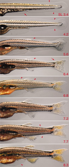
Larval fin fold and fin fold resorption. Multiple individuals are shown (standard length [SL] at lower right). 3.4, Initial shapes of fin fold major lobe (arrowheads) and minor lobe (arrow); 4.2, a constriction is evident at the posterior tail (arrowhead); 5.6, a bulge is evident in the dorsal fin fold above the dorsal fin mesenchymal condensation (arrowhead); 6.4, a notch posterior to the dorsal fin indicates early fin fold resorption (arrowhead) and a bulge is evident over the developing supranotochordal fin rays (arrow); 6.8, resorption continues both anterior and posterior to the dorsal fin (arrowheads); 7.7, resorption occurs in an increasingly posterior zone along the tail (arrowheads); 8.3, early resorption of the minor lobe is revealed by flattening of its ventral posterior margin (arrowhead; also see Fig. 22). |