
Fig. 1
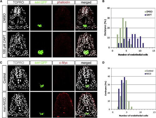
Notch Signaling Negatively Regulates the Number of Endothelial Cells during Zebrafish Development (A) Transverse sections of 18 hpf embryos, DMSO- or DAPT-treated, visualized for TOPRO (white), kdrl:GFP (green), and phalloidin (red). (B) Quantification of endothelial nuclei per focal plane in DMSO-treated (n = 30) or DAPT-treated (n = 44) embryos. (C) Transverse sections of 18 hpf phenotypic wild-type siblings and Tg(hsp70l:Gal4)kca4;Tg(UAS:myc-NICD)kca3 embryos visualized for TOPRO (white), kdrl:GFP (green), and c-Myc as a surrogate measure for NICD expression (red). (D) Quantification of endothelial nuclei per focal plane in phenotypic wild-type (n = 31) and NICD-overexpressing (n = 28) embryos. Embryos treated with DAPT contained 6.66 (s = 2.42) endothelial nuclei per section, whereas those treated with DMSO had 4.03 (s = 1.27) endothelial nuclei (ANOVA, p < 10-6). Conversely, embryos overexpressing the NICD contained 3.14 (s = 0.80) endothelial nuclei, in comparison to 4.42 (s = 1.09) nuclei per section in phenotypic wild-type siblings (ANOVA, p < 10-5), suggesting that Notch signaling negatively regulates endothelial cell number. |
| Gene: | |
|---|---|
| Fish: | |
| Conditions: | |
| Anatomical Term: | |
| Stage: | 14-19 somites |