
Fig. 1
- ID
- ZDB-FIG-090710-51
- Publication
- Yabe et al., 2009 - The maternal-effect gene cellular island encodes aurora B kinase and is essential for furrow formation in the early zebrafish embryo
- Other Figures
- All Figure Page
- Back to All Figure Page
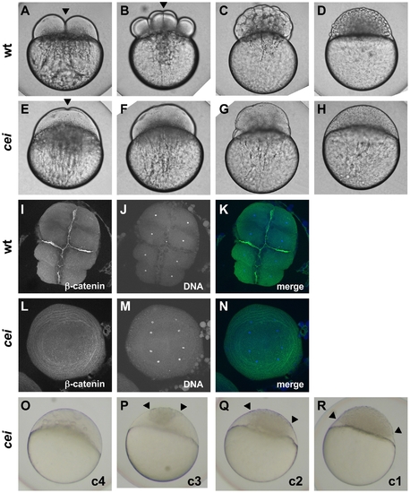
Embryos from cei homozygous females exhibit defects in cytokinesis. Cytokinesis defects in live (A–H, O–R) and fixed (I–N) wild-type (A–D, I–K) and cei mutant (E–H, L–R) embryos. (A–H) Side views of live wild-type (A–D) and maternally mutant cei (E–H) embryos at time points equivalent to (in wild-type embryos) the 2-cell (A,E), 8-cell (B,F), 64-cell (C,G) and 1,000-cell (D,H) stages. Maternally mutant cei embryos exhibit a rudimentary furrow (arrowhead in E) and form syncytial embryos (H). (I–N) Animal views of fixed wild-type (I–K) and maternally mutant cei (L–N) embryos, labeled with an antibody against β-catenin, a component of cell adhesion junctions present in mature furrows (I,L) and the DNA dye DAPI (J,M). Merged images shown in (K,N). (O–R) Side views of live embryos at the 1,000 cell stage, showing the phenotypic range of maternally mutant cei embryos. Embryos exhibit various degrees of aberrant syncytium formation. Images are representative for the categories presented in Table 1: C4 (most severe – O), C3 (P), C2 (Q), C1 (least severe – R). Arrowheads show the limits of a single cellularized region that typically sits atop the syncytial region. |
| Fish: | |
|---|---|
| Observed In: | |
| Stage Range: | 8-cell to 1k-cell |