
FIGURE
Fig. S3
- ID
- ZDB-FIG-090617-45
- Publication
- Bae et al., 2009 - Anatomy of zebrafish cerebellum and screen for mutations affecting its development
- Other Figures
- All Figure Page
- Back to All Figure Page
Fig. S3
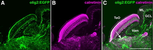
olig2:EGFP+ axons and calretinin-immunoreactivity (CR-ir) in pretectal region. The brain of adult Tg(olig2:EGFP) fish were stained with anti-EGFP (green, A) and anti-calretinin antibodies (magenta, B). (C) Superimposed view of A and B. Sagittal section. The pretectal region is indicated by arrow. Note that both olig2:EGFP+ fibers and CR-ir+ neurons/fibers were detected in the preoptic regions, and also broadly in other regions of the brain. The abbreviations are described in Fig. 1. Scale bar: 500 μm. |
Expression Data
Expression Detail
Antibody Labeling
Phenotype Data
Phenotype Detail
Acknowledgments
This image is the copyrighted work of the attributed author or publisher, and
ZFIN has permission only to display this image to its users.
Additional permissions should be obtained from the applicable author or publisher of the image.
Reprinted from Developmental Biology, 330(2), Bae, Y.K., Kani, S., Shimizu, T., Tanabe, K., Nojima, H., Kimura, Y., Higashijima, S.I., and Hibi, M., Anatomy of zebrafish cerebellum and screen for mutations affecting its development, 406-426, Copyright (2009) with permission from Elsevier. Full text @ Dev. Biol.