
FIGURE
Fig. S1
- ID
- ZDB-FIG-090617-43
- Publication
- Bae et al., 2009 - Anatomy of zebrafish cerebellum and screen for mutations affecting its development
- Other Figures
- All Figure Page
- Back to All Figure Page
Fig. S1
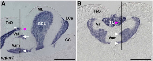
Expression of vglut1 (slc17a7). In situ hybridization of sagittal (A, identical to Fig. 3B) and cross sections (B) of the adult cerebellum. Lines in A and B indicate the corresponding positions of section B and A, respectively. Magenta arrowheads indicate vglut1 expression in the torus longitudinalis (TL), and white arrows and arrowheads indicate its expression in the granule cell layer (GCL). The abbreviations are described in Fig. 1. TL, torus longitudinalis. Scale bars: 500 μm. |
Expression Data
Expression Detail
Antibody Labeling
Phenotype Data
Phenotype Detail
Acknowledgments
This image is the copyrighted work of the attributed author or publisher, and
ZFIN has permission only to display this image to its users.
Additional permissions should be obtained from the applicable author or publisher of the image.
Reprinted from Developmental Biology, 330(2), Bae, Y.K., Kani, S., Shimizu, T., Tanabe, K., Nojima, H., Kimura, Y., Higashijima, S.I., and Hibi, M., Anatomy of zebrafish cerebellum and screen for mutations affecting its development, 406-426, Copyright (2009) with permission from Elsevier. Full text @ Dev. Biol.