
Fig. 2
- ID
- ZDB-FIG-090511-16
- Publication
- Willett et al., 1997 - Expression of zebrafish rag genes during early development identifies the thymus
- Other Figures
- All Figure Page
- Back to All Figure Page
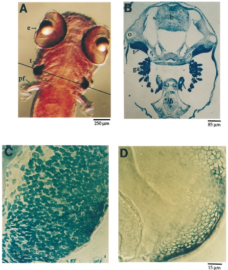
Sections of fish hybridized to rag1 probe identify the zebrafish thymus. (A) A dorsal view of a 1-week-old fish hybridized with the rag1 antisense probe. The thymus (t) is clearly visible. A line across the posterior head indicates the plane of the section shown in B. The section in B has been stained with toluidine blue and shows the location of typically lymphoid organs in the head identified as the bilaterally symmetric thymus (t). The thymus lies just ventral to the otic vesicle (o) in the plane of the heart (h). C and D are sequential sections of the fish shown in A; in addition, C has been stained with toluidine blue which shows that this organ is filled with small cells possessing large nuclei, typical of thymocytes. Comparison of B, C, and D shows that the organ having a typically lymphoid appearance in B hybridizes to the rag1 probe. b, brain; e, eye; ga, gill arch; h, heart; o, otic vesicle; pf, pectoral fin; t, thymus. |
| Gene: | |
|---|---|
| Fish: | |
| Anatomical Term: | |
| Stage: | Days 7-13 |
Reprinted from Developmental Biology, 182(2), Willett, C.E., Zapata, A.G., Hopkins, N.A., and Steiner, L.A., Expression of zebrafish rag genes during early development identifies the thymus, 331-341, Copyright (1997) with permission from Elsevier. Full text @ Dev. Biol.